Brain Mapping in the Frequency Domain for Autism Children:
A Magnetoencephalographic Study with pT-Transcranial Brain Stimulation
Photios A. Anninos1, Athanasios Chatzimichael2, Adam Adamopoulos1, Athanasia Kotini1, Nicolaos Tsagas3
2.Department of Paediatrics, University Hospital of Alexandroupoli, Democritus University of Thrace, Alexandroupoli, Greece.
3.Department of Electrical Engineering, Polytechnic School, Democritus University of Thrace, Xanthi, Greece.
Citation : Anninos P, Chatzimichael A, Adamopoulos A, Kotini A, Tsagas N. Brain Mapping in the Frequency Domain for Autism Children: A Magnetoencephalographic Study with pT-Transcranial Brain Stimulation. Clin Res Neurol 2018;1(2): 1-4.
The technique of transcranial magnetic brain stimulation (TMS) has investigational, indicative, and beneficial potential. Anninos and Tsagas [1] created and designed a pico Tesla-TMS electronic tool that is a modified helmet including up to 122 coils so as to cover up the right and left temporal, right and left parietal, frontal, vertex, and occipital regions of each patient. It generates pT-TMS range modulations of magnetic flux in the patient's alpha rhythm (8-13 Hz) and creates a square wave so as to look like the firing activity of the brain neurons.
In this commentary, we provide brain maps after the application of fast Fourier transform (FFT) as a continuation of our formerly published studies [2-4].
The method of the research has been published in detail in our previous work [2-4]. Eight autistic children (five boys and three girls, with ages ranged from 5 to 12 years, mean ± SD: 8.5 ± 2.3) were included in the study. Magnetoencephalographic (MEG) measurements were performed using a whole-head 122-channel MEG system (Neuromag-122, Neuromag Ltd., Helsinki, Finland) [2-4] in an electromagnetically shielded room. The Research Committee of our University approved the study. Our laboratory developed a software program that identifies the amplitude of the primary dominant frequency of the power spectra of the MEG after the application of FFT. Afterward, we are interesting for the alpha-frequency (8-13 Hz) for calibration of the electronic device and the (2-7 Hz) frequencies for the analysis to obtain the principal dominant frequencies and the power spectra of the MEG [1-4].
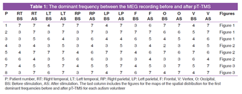
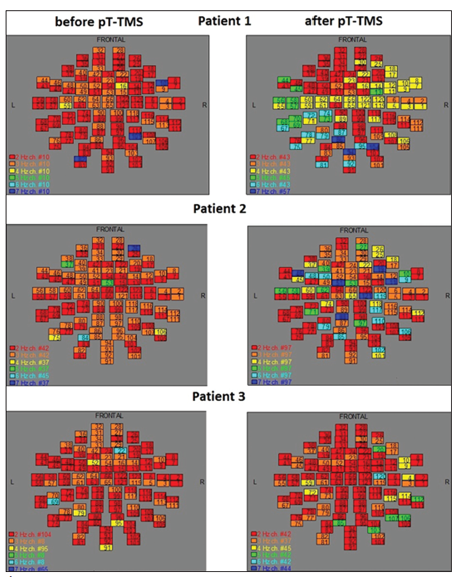
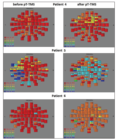
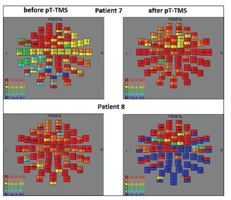
We used the FFT algorithm to obtain the maps of the power spectra. Different colors in the maps correspond to different dominant frequencies. The numbers in the map squares represent the 122 MEG channels.
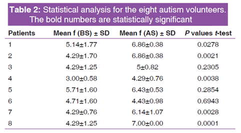
In Table 1, the BS and AS represent the effect prior and after the pT-TMS for each autism volunteer. Table 2 shows the statistics for the autism patients using unpaired t-test. The results were significant at 5 patients (62.5%). We see that five of eight patients have got improvement according to the statistical analysis of Table 2.
Figures 1-3 represent the maps of the application of FFT on MEG data before and after pT-TMS for each autism patient.
This means of pT-TMS has prospective to be a significant non-invasive secure method in managing autism patients while extra investigations with a larger number of patients are suggested before have firm conclusions.
REFERENCES
- Anninos PA, Tsagas N. Electronic Apparatus for Treating Epileptic Individuals. USA patent 5453072; 1995.
- Anninos P, Kotini A, Adamopoulos A, Tsagas N. MEG and pico-Tesla-TMS in patients with dystonia or autism (Commentary). J Neurol Neuro Toxicol 2017;1:23-59.
- Anninos P, Chatzimichael A, Adamopoulos A, Kotini A, Tsagas N. Autism disorder and pico-Tesla TMS. Arch Paediatr Dev Pathol 2017;1:1008.
- Anninos P, Chatzimichael A, Adamopoulos A, Kotini A, Tsagas N. A combined study of MEG and pico-Tesla TMS on children with autism disorder. J Integr Neurosci 2016;15:497-513.